Histology slides of exocrine and endocrine glands This part of this article will show you the exocrine and endocrine histology slides with their identification points Make sure you know the structures and function of different types of acinus like – mucous acini, serous acini, and seromucous aciniThe term "endocrine" implies secretion into the internal milieu of a multicellular organism In contrast to exocrine tissues, where the secretory products are discharged into the external space the outer surface of the body, mucosal surfaces, duct systems the endocrine organs and cells secrete their products into the vascular system This is achieved by the bicarbonate secretions of the pancreas Exocrine Pancreas – The Functional Unit The functional unit of the exocrine pancreas includes the acinus and its duct system The word acinus is from the Latin term for "berry in a cluster" These acinar cells are specialised in enzyme synthesis, storage and secretion

Olanzapine Induced Biochemical And Histopathological Changes After Its Chronic Administration In Rats Sciencedirect
Exocrine pancreas histology labeled
Exocrine pancreas histology labeled-The pancreas is lumpy and glandular It comprises a head, neck, body, and tail, which points towards the spleen on the left side of the body The duodenum wraps around the head of the pancreas Exocrine Tissues The pancreatic tissue primarily comprises acini, which are clusters of secretory cells Visible as a circle of dark purplestaining226 Pancreas Exocrine Pancreas View Virtual EM Slide In this low power electron micrograph, observe the organization of the acini, composed of acinar cells Within the acinar cells you will see the basal rough endoplasmic reticulum and the numerous secretory granules in the apical region of the cells, facing the small lumen of the acinus


Labeled Pancreas Histology
In some animals, two ducts enter the duodenum rather than a single duct In some species, the main pancreatic duct fuses with the common bile duct just before its entry into the duodenum Additional information on microscopic anatomy of the pancreas can be found in the section on Histology of the PancreasThe pancreas is formed from two basic tissue types termed the exocrine and endocrine pancreas Although the exocrine and endocrine pancreas share a similar anatomic location, for all intensive purposes they are histologically and physiologically distinct So too are the pathological processes which affect themThe pancreas is a 2in1 organ an exocrine and endocrine gland It is essential for digestion and the carbohydrate metabolism Thus, a loss in pancreatic function leads to severe clinical symptoms In this article, you will get a compact overview of the structure, functions, and diseases of the pancreas Macroscopic Anatomy of the Pancreas
Anatomy Parotid gland Largest salivary gland (15 30 g), 6 x 3 cm It is wrapped around the mandibular ramus, has broad superficial lobe and smaller deeper lobe, with facial nerve usually between both lobes Provides only 25% of the total salivary volume but on stimulation, the parotid secretion rises to 50%Note also that the acinar cells are clearly polarized their basal cytoplasm (periphery of acinus) is quite basophilic, owing to large accumulations of RER, whereas theirPancreas histology ,pancreas histology labeled ,histology pancreas ,histology of pancreas ,exocrine pancreas histology ,normal pancreas histology ,pancreas histology slide ,histology of the pancreas ,pancreas gland histology ,pancreas histology slides ,pancreas location ,islets of langerhans ,pancreas definition ,where is the pancreas located ,pancreas anatomy ,pancrease ,pancreatic
The pancreas is functionally and histologically divided into exocrine and endocrine components In the exocrine component , there are zymogenic cells that are pyramidal and form the serous acini The gland is also lobulated by fibrous septae that contains general neurovascular structures as well as specialized nerves called Pacinian corpuscles4050 acinar cells form a round to oval acinus;By Geoff Meyer All body organs are made up of the four basic tissues epithelium, connective tissue, muscle and nervous tissue In this course, students are presented detailed descriptions of the histological structure and cellular specializations of organs and organ systems of the body accompanied by high resolution images of each tissue and
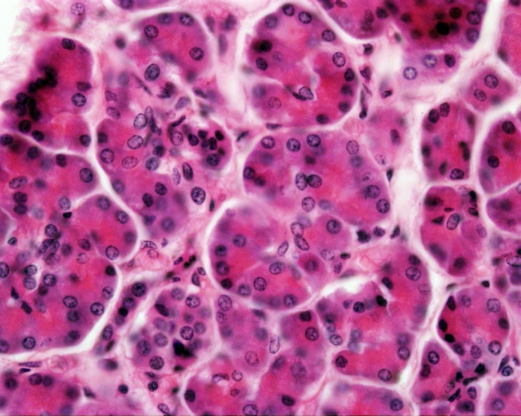


File Pancreas Histology 102 Jpg Embryology


Liver Gall Bladder Pancreas
Histology in the Pancreas and MRI Imaging Biomarkers Temel Tirkes, Indiana University Organoid and Pancreas on Chip Alex Kleger, MD, Ulm University 3D Imaging Study and Mapped Changes in Innervation in Type 2 Diabetes Sarah Stanley, MBBCh, PhD, Mount Sinai Pancreas Slices to Study Interactions between Immune Cells and Islets The functional unit of the exocrine pancreas is the acinus (pl acini) Acini are organized collections of secretory cells that surround a central lumen Acinar cells have peripherally located nuclei and abundant granular cytoplasm and secrete a variety of active enzymes and inactive proenzymes into the acinar lumen ( Figure 113 )Lumen of acinus is occupied by 3 or 4 centroacinar cells, which form the beginning of the duct system;
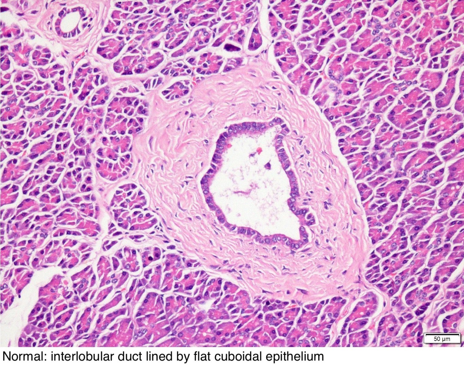


Pathology Outlines Panin
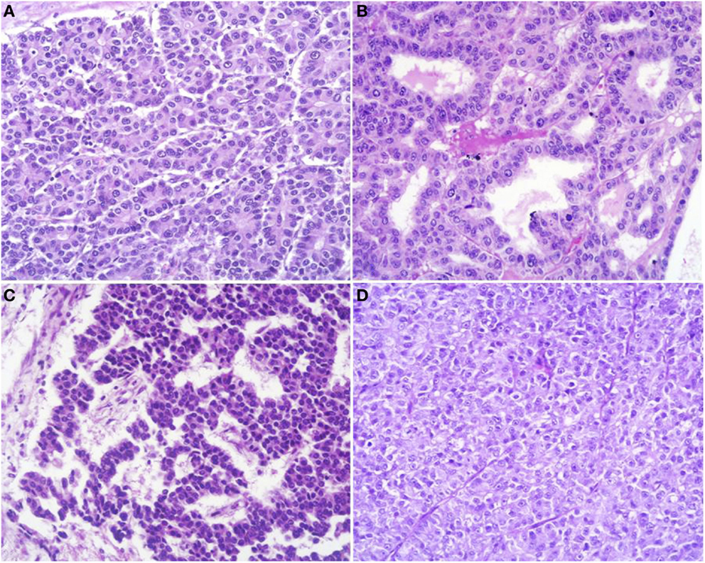


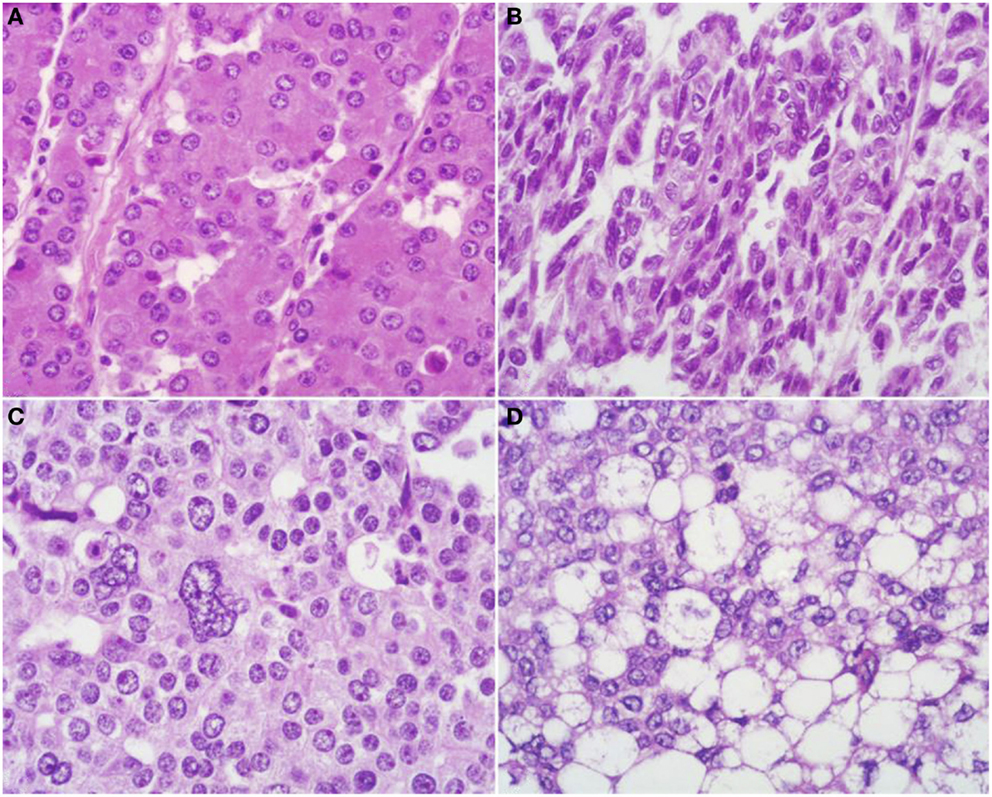
Frontiers Acinar Cell Carcinoma Of The Pancreas Overview Of Clinicopathologic Features And Insights Into The Molecular Pathology Medicine
View extensive video descriptions of all the histological features of human organs within the human organ systems cardiovascular system, respiratory system, endocrine system, male reproductive system, female reproductive system, urinary system, lymphoid organs or lymphatic system, oral cavity and the digestive system, histology of the liver, gallbladder and pancreasPancreas, Exocrine / anatomy & histology Pancreas, Exocrine / metabolism Salivary Glands / anatomy & histology Salivary Glands / metabolism Substances Electrolytes Grant support P01 HD/HD/NICHD NIH HHS/United States R01 DE/DE/NIDCR NIH HHS/United StatesThe pancreas is located posterior to the stomach and next to the duodenum The pancreas functions as both an exocrine and endocrine gland The exocrine function of the pancreas is essential for digestion as it produces many of the enzymes that break down the protein, carbohydrates, and fats in digestible foods


Liver Gall Bladder Pancreas
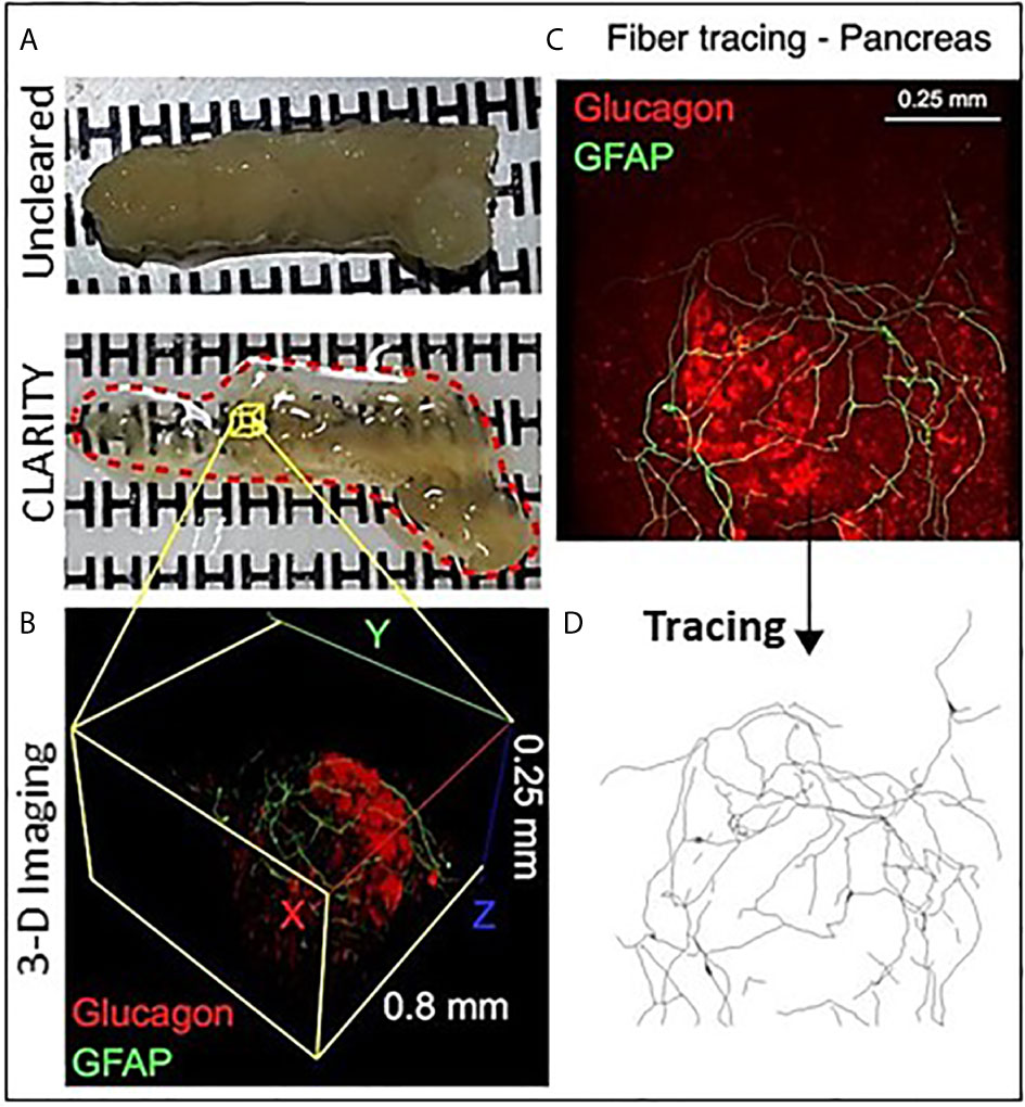


Frontiers Pancreas Optical Clearing And 3 D Microscopy In Health And Diabetes Endocrinology
Pancreas Has both exocrine and endocrine functions • Most of the pancreas comprises exocrine cells, called acini, that secrete digestive enzymes; 23 Anatomy, Histology, & Embryology of the Pancreas *P ancreas is secondary retroperitoneal, with the exception of the tail, the foregut Anatomy of Pancreas Location Within the curve of the duodenum, located in the epigastric and left hypochondriac regions Surface Projection Surface projection of is different depending on the part of it, and will be entailed laterPancreas is an exocrine organ The exocrine function involves producing digestive enzymes released into the small intestine We make more digestive enzymes than insulin so most of the cells are acinar cells that produce digestive enzymes The pancreatic isles, also called isles of Langerhan, produce insulin It is very difficult to


Pancreas Histology
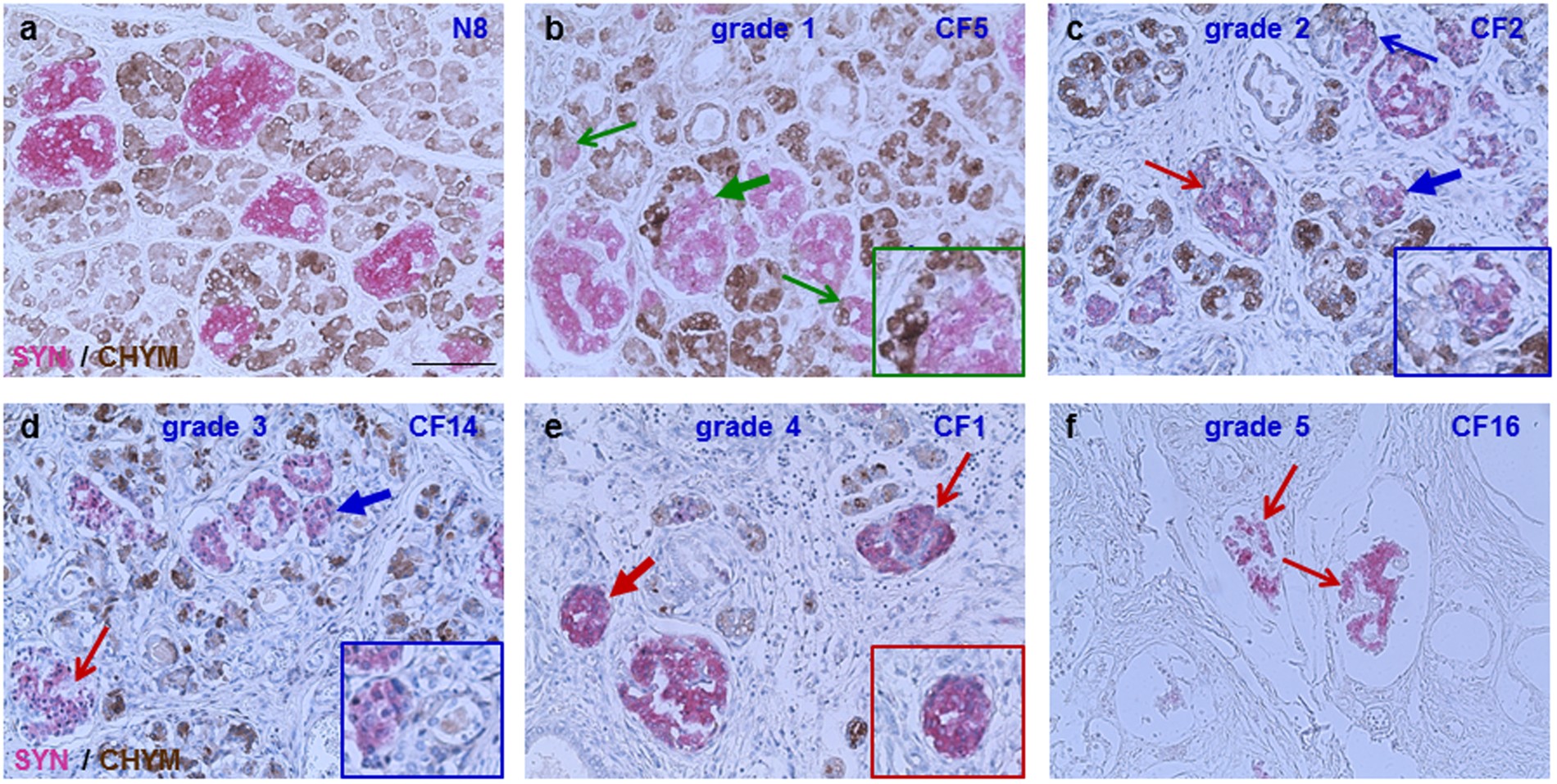


Structural Abnormalities In Islets From Very Young Children With Cystic Fibrosis May Contribute To Cystic Fibrosis Related Diabetes Scientific Reports
Pancreata from rodents and humans share many histological and functional similarities, but also display some potentially important differences ()The pancreas is invested by a variably thin connective tissue capsule and divided into lobules, which are formed from dense accumulations of exocrine glands that often surround the isletsIndividual lobules are defined by5 VHL and the pancreas VHL increases risk of several types of pancreatic masses and cysts Mass is solid Cyst is fluid filled Most will be benign One type of mass seen is a pancreatic neuroendocrine tumor Pancreatic Neuroendocrine Tumors (PNETs) 1015% of all PNETs are associated with genetic syndromesPathology Outlines Anatomy & histolog 309 histology pancreas stock photos, vectors, and illustrations are available royaltyfree See histology pancreas stock video clips of 4 islet of langerhans pancreas cells endocrine cells islet cells exocrine diabetes pattern langerhans pancreas beta cells islet cell diabetes microscope


Pancreatic Histology Exocrine Tissue


Pancreatic Ductal Deletion Of Hnf1b Disrupts Exocrine Homeostasis Leads To Pancreatitis And Facilitates Tumorigenesis Cellular And Molecular Gastroenterology And Hepatology
Exocrine Pancreas The exocrine part of the pancreas has closely packed serous acini, similar to those of the digestive glands It secretes an enzyme rich alkaline fluid into the duodenum via the pancreatic duct The alkaline pH is due to the presence of bicarbonate ions, and helps to neutralise the acid chyme from the stomach, as it enters the by anatomylearner In pancreas histology of animal, you will find the both endocrine and exocrine parts The exocrine part forms the major portion of pancreas and consists of closely packed serous acini with zymogenic cells Again, the endocrine part consists of pancreatic islets of Langerhans which is located within the masses of serous acini The pancreas is a twoheaded organ, not only in origin but also in function In origin, the pancreas develops from two separate primordia In function, the organ has both endocrine function in relation to regulating blood glucose (and also other hormone secretions) and gastrointestinal function as an exocrine (digestive) organ, see exocrine pancreas


Anatomy And Histology Of The Pancreas Pancreapedia


Digestive The Histology Guide
Labeled sensory neurons (about 2,700 cells) were found bilaterally in the dorsal root ganglia T3 to L5, chiefly in T10 to L1 Some differences were found in the localization of labeled postganglionic and sensory neurons between the two portions of the pancreas Injection in to the splenic portion revealed more labeled neurons in ganglia on the left side, while injection into the duodenal portion gave rise to a greater number of labeledPDFANATOMY AND HISTOLOGY OF THE PANCREAS The pancreas is a particularly important organ from the point of view of human medicine because it suffers from two important diseases diabetes mellitus and pancreatic cancer, Although the exocrine and endocrine pancreas share a similar anatomic location, Four main cell types exist in the isletsThe ducts that drain them secrete alkaline fluid • Clusters of endocrine cells, called Islets of Langerhans, secrete hormones that regulate blood glucose metabolism Key Anatomical & Histological Features The pancreas is lumpy and


2


Morphological Histological And Ultrastructural Studies On The Exocrine Pancreas Of Goose Sciencedirect
Exocrine pancreas, the portion of the pancreas that makes and secretes digestive enzymes into the duodenum This includes acinar and duct cells with associated connective tissue, vessels, and nerves The exocrine components comprise more than 95% of the pancreatic massExocrine pancreas Secretory units pancreatic acini Cells acinar cells, centroacinar cells Products peptidases, lipases, amylolytic enzymes, nucleolytic enzymes Endocrine pancreas Secretory units islets of Langerhans Cells A (alpha), B (beta), D (delta), PP (pancreatic polypeptide) cells Histology of the exocrine pancreas Pietro M Motta, Corresponding Author Department of Anatomy, University of Rome La Sapienza, I Rome, Italy Department of Anatomy, University of Rome La Sapienza, Via A Borelli 50, I Rome, ItalySearch for more papers by this author


File Pancreas Histology 103 Jpg Embryology


Chapter 15 Page 7 Histologyolm 4 0
The pancreas is a large, relatively flat, whitetopinkcolored organ that develops from an outgrowth of the primitive foregut It is a combined endocrine and exocrine gland in close association with the upper duodenum During development, exocrine and endocrine elements are both derived from differentiation of duct epithelium HMB Pancreas Histology Liver Histology Gall Bladder Histology Renal System Histology exocrine pancreas consists of tubuloacinar glands single layer of pyramidal shaped cells forms the secretory acini (cells contain zymogen granules) Secretory duct pathway Intercalated Duct;The pancreas develops from two outgrowths of the foregut *The ventral diverticulum gives rise to the common bile duct, gallbladder, liver and the ventral pancreatic anlage that becomes a portion of the head of the pancreas with its duct system including the uncinate portion of the pancreas *The dorsal pancreatic anlage gives rise to a portion of the head, the body, and tail of the pancreas


Labeled Pancreas Histology


Pancreatic Histology Endocrine Tissue
Key facts about the histology of the pancreas;The mandate for this chapter is to review the anatomy and histology of the pancreas, an endocrine organ that makes and secretes hormones into the blood to control energy metabolism and storage throughout the body The mandate for this chapter is to review the anatomy and histology of the pancreas The pancreas (meaning all flesh) lies in the upper abdomen behind The morphology of the exocrine secretory unit of the pancreas, ie the pancreatic acinus, is reviewed The histological features of the acini and their relation with the duct system are described The acinar three‐dimensional architecture was studied by means of different ultrastructural techniques, some of which are complementary


Deletion Of Xiap Reduces The Severity Of Acute Pancreatitis Via Regulation Of Cell Death And Nuclear Factor Kb Activity Cell Death Disease


Scielo Brasil Comparative Evaluation Of Pancreatic Histopathology Of Rats Treated With Olanzapine Risperidone And Streptozocin Comparative Evaluation Of Pancreatic Histopathology Of Rats Treated With Olanzapine Risperidone And Streptozocin
Exocrine vs Endocrine Fundamentally, the difference between exocrine and endocrine is exocrine secretes (via ducts) to an external surface or body cavity endocrine secretes within an enclosed system eg the bloodstream As the acini of the pancreas secrete into a network of ducts, this gives this organ an exocrine functionA brief review of the normal histology of the pancreas, as presented by the URMC Pathology IT ProgramThe pancreas is an organ of the digestive system and endocrine system of vertebratesIn humans, it is located in the abdomen behind the stomach and functions as a glandThe pancreas has both an endocrine and a digestive exocrine function As an endocrine gland, it functions mostly to regulate blood sugar levels, secreting the hormones insulin, glucagon, somatostatin, and pancreatic
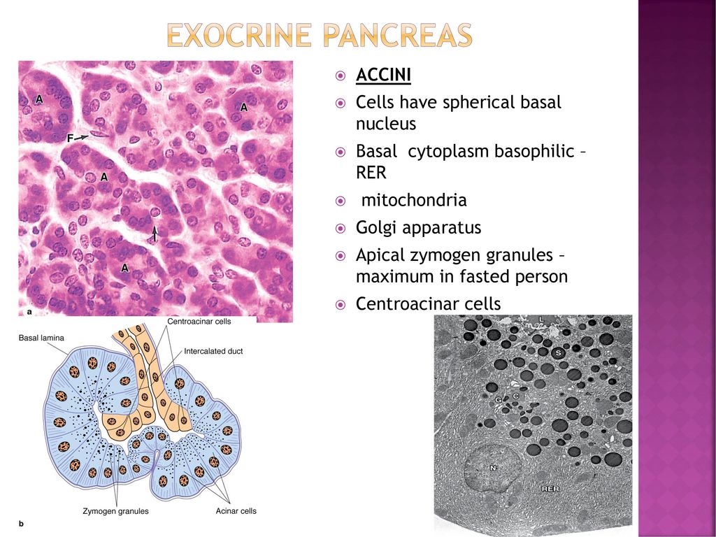


Histology Of Pancreas Ppt Download


P2ry1 Alk3 Expressing Cells Within The Adult Human Exocrine Pancreas Are Bmp 7 Expandable And Exhibit Progenitor Like Characteristics Cell Reports
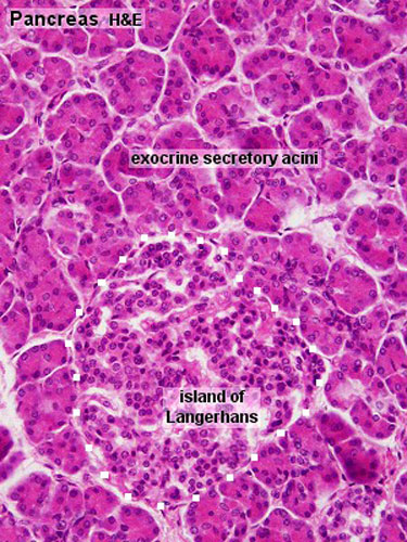
At the histological level the pancreas is made up of compound glands in "bunch of grapes" fashion The pancreas has an exocrine and endocrine component The exocrine compnent is demonstrated above in 3D with acini in "cluster of grapes" formation subtended by a duct Courtesy Ashley Davidoff MD a06 Exocrine pancreas Using the high dry objective (6, H&E 40x, 40xlabeled ), study the structure of several acini Note that the acinar cells are pyramidal in shape;The pancreas is a long, slender organ, most of which is located posterior to the bottom half of the stomach (Figure 1791)Although it is primarily an exocrine gland, secreting a variety of digestive enzymes, the pancreas also has endocrine cells Its pancreatic islets—clusters of cells formerly known as the islets of Langerhans—secrete the hormones glucagon, insulin, somatostatin, and


Pin By Tracey Bradfield On A P Pancreas Endocrine Endocrine System


Histoquarterly Pancreas Histology Blog Medical School Stuff Histology Slides Endocrine System
Describe the exocrine pancreas histologically composed of tubuloacinar gland that produces about 10 ml of bicarbrich fluid containing digestive proenzymes and enzymes;Histology @ Yale Slide List Pancreas Pancreas This image shows the two main functional domains of the pancreas The endocrine pancreas that secretes insulin and glucogon is more lightly stained and its cells cluster to form the Islets of Langerhans The rest of the image consists of the exocrine pancreas that produces several enzymesThe pancreas is a glandular organ located in the retroperitoneum between the duodenal curvature and the splenic hilum It has both exocrine and endocrine functions and is anatomically classified into four parts the head, neck, body, and tail


Human Pancreatic Afferent And Efferent Nerves Mapping And 3 D Illustration Of Exocrine Endocrine And Adipose Innervation American Journal Of Physiology Gastrointestinal And Liver Physiology


Pancreatic Histology Acinar Cells Produce Pancreatic Juice And Make Up The Bulk Of The Pancreas Studying Medicine Histology Slides Medical Laboratory Science
Intralobular Duct (low columnar or cuboidal epithelium, nonstriated) Interlobular Duct (columnar epithelium goblet cells) Main Pancreatic The exocrine secretory part consists of serous acini (known as pancreatic acini), whereas the islets of Langerhans form the endocrine part The major part of pancreas corresponds to the exocrine part (99 % in humans) The serous exocrine cells show a rounded nucleus and dark cytoplasm Histology serous ductal / acinar (exocrine cells) gland with tubuloalveolar arrangement with interspersed islets of Langerhans (endocrine cells) Terminology Anatomically, from right to left, the pancreas is divided into the caput (head), collum (neck), corpus (body) and cauda (tail) Pancreatic secretion is drained by the main pancreatic duct


Biology 2404 A P Basics


Morphology Of Salivary And Lacrimal Glands Intechopen


Dictionary Normal Pancreas The Human Protein Atlas


Pancreas Histology Labeled
:watermark(/images/watermark_5000_10percent.png,0,0,0):watermark(/images/logo_url.png,-10,-10,0):format(jpeg)/images/overview_image/1059/SXYoNKo9I26Aroj0YPbuHg_pancreatic-duct-system_english.jpg)


Pancreas Histology Exocrine Endocrine Parts Function Kenhub


Histology Endocrine System Pancreas


Digestive The Histology Guide


Histology Of Pancreas Youtube
:watermark(/images/watermark_5000_10percent.png,0,0,0):watermark(/images/logo_url.png,-10,-10,0):format(jpeg)/images/overview_image/1881/CPsIC67xuz6pLt6JhAM6Wg_pancreas-histology_english.jpg)


Pancreas Histology Exocrine Endocrine Parts Function Kenhub


Pancreas Histology Identifying Features With Labeled Slide Images Anatomylearner The Place To Learn Veterinary Anatomy Online


Pancreas Wikipedia


Rabbit Histology Pancreas Rabbit Labels Histology Slide


Histoquarterly Pancreas Histology Blog


Anatomy And Histology Of The Pancreas Pancreapedia
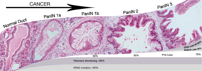


Surgical And Molecular Pathology Of Pancreatic Neoplasms Diagnostic Pathology Full Text


Anatomy And Histology Of The Pancreas Pancreapedia
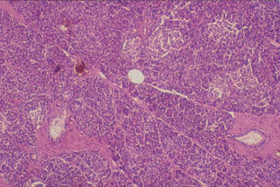


Histology World Histology Fact Sheet Pancreas
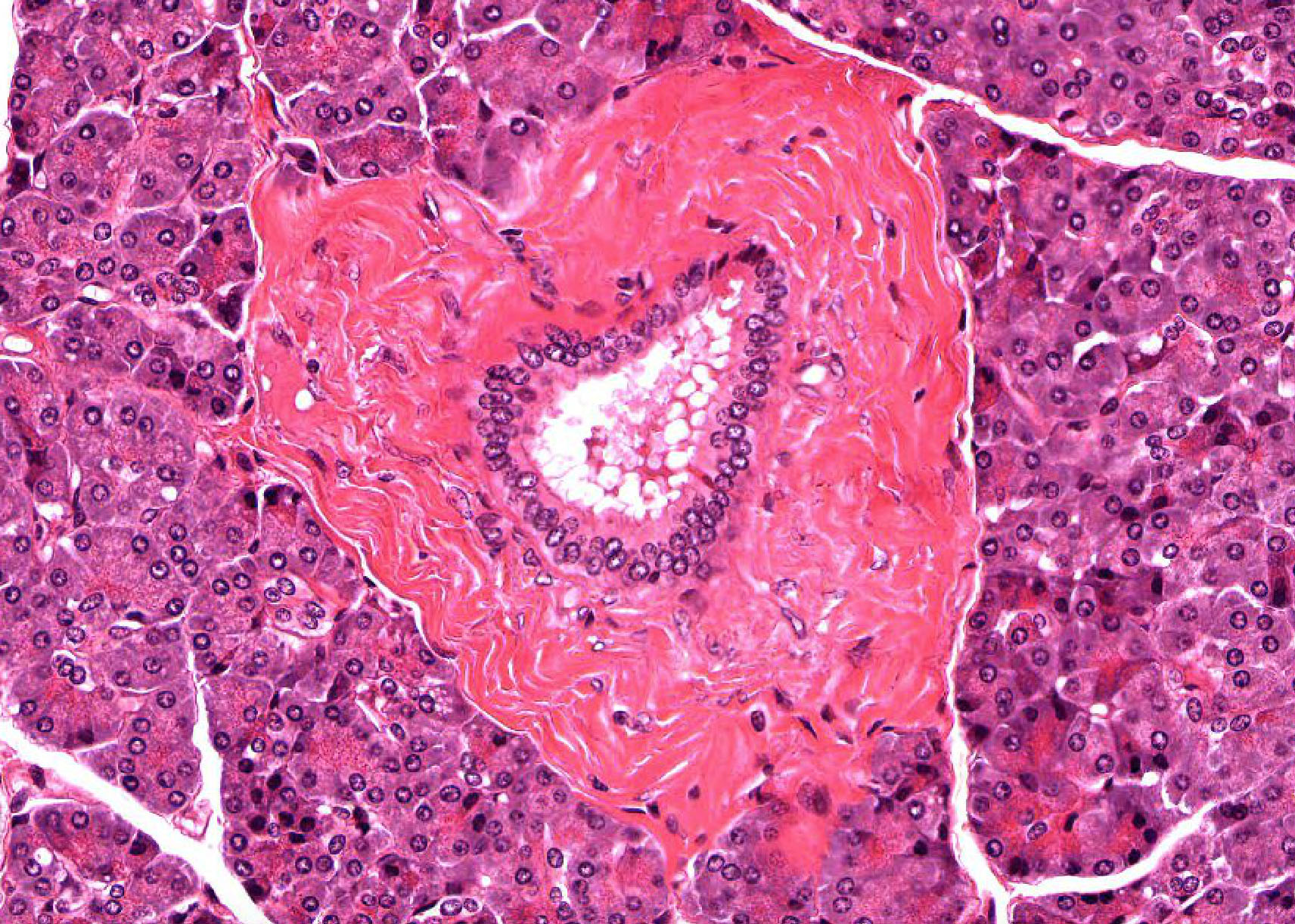


Pancreas Histology


Olanzapine Induced Biochemical And Histopathological Changes After Its Chronic Administration In Rats Sciencedirect


Structure Of The Pancreatic Acini A The Pancreatic Acini Formed From Download Scientific Diagram


Digestive


2


Pancreas Histology
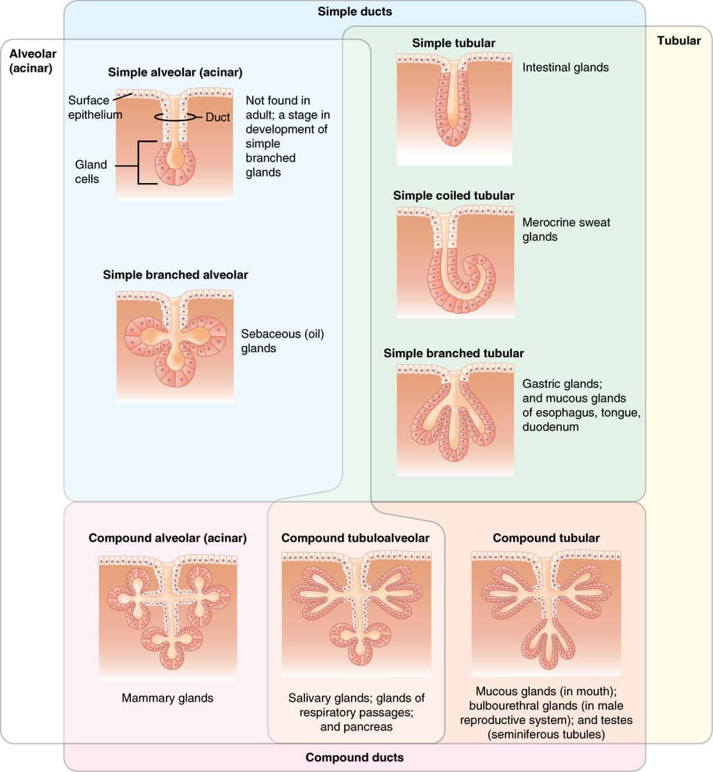


Structure Of Glands Exocrine Endocrine Histology Teachmephysiology


Pancreatic Histology Of Pdx1 Mutants A D Gut Region Morphology In Download Scientific Diagram


1


Light Micrograph Of The Exocrine Pancreas At High Magnification


Liver And Pancreas


Pancreatic Histology Exocrine Tissue
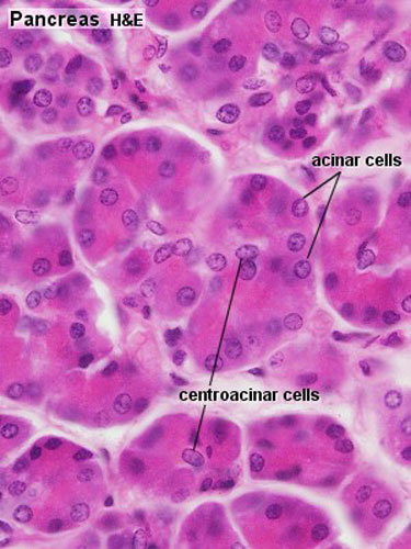


File Pancreas Histology 002 Jpg Embryology


Pancreas Histology Flashcards Quizlet


Histology Endocrine System Pancreas
:watermark(/images/watermark_only.png,0,0,0):watermark(/images/logo_url.png,-10,-10,0):format(jpeg)/images/anatomy_term/pancreatic-acinar-cells/86g1HQPABGT8iQy0zPdrpg_Exocrine_cell_of_pancreas.png)


Pancreas Histology Exocrine Endocrine Parts Function Kenhub


Pancreas Histology Flashcards Quizlet


Pathology Of Endocrine Pancreas And Complications Of Diabetes Flashcards Quizlet


Endocrine Pancreas


Basic Histology Exocrine Pancreas


2


Pancreas Histology


Pancreas Anatomy Histology Histology Flashcards Draw It To Know It


Pancreas Histology Flashcards Quizlet
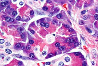


Histology Of The Pancreas Springerlink


Pancreas Slide
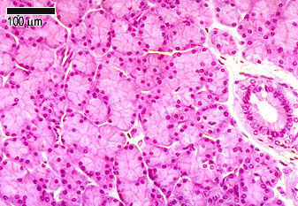


Glandular Tissue The Histology Guide
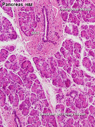


Gastrointestinal Tract Pancreas Histology Embryology


Combination Of Obestatin And Bone Marrow Mesenchymal Stem Cells Prevents Aggravation Of Endocrine Pancreatic Damage In Type Ii Diabetic Rats Abstract Europe Pmc


2


Histology Website Resource Ha37 Pancreas Cat


Anatomy And Histology Of The Pancreas Pancreapedia


2


Ha 235 Histology Accessory Digestive Glands
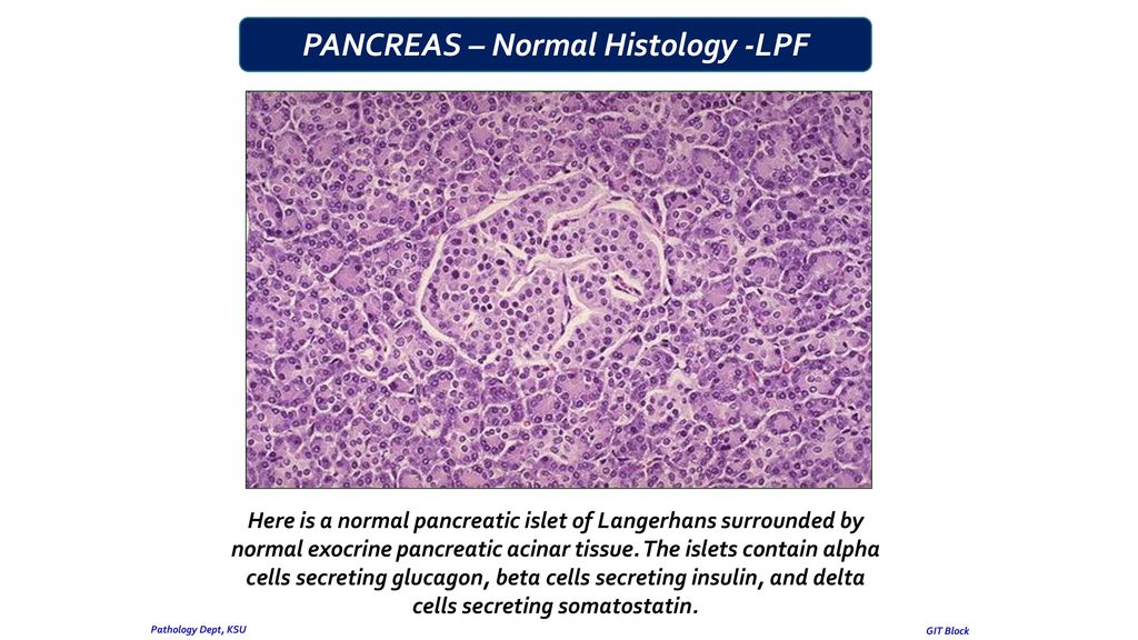


Pancreas Pathology Dept Ksu Git Block Ppt Download


Histology Panosundaki Pin


Structure Of The Pancreatic Acini A The Pancreatic Acini Formed From Download Scientific Diagram


Acvim Consensus Statement On Pancreatitis In Cats Forman 21 Journal Of Veterinary Internal Medicine Wiley Online Library


Pancreatic Histology Exocrine Tissue


Pancreatic Histology Exocrine Tissue


2


Pancreas Slide Wsu 2 060


Deletion Of Dicer Alters Histology In Exocrine Pancreas A Download Scientific Diagram


2
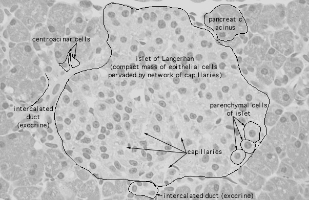


Hls Endocrine System Pancreas Islets Of Langerhans High Mag Labeled


Gastrointestinal Tract Pancreas Histology Embryology


Spontaneous Induction Of Murine Pancreatic Intraepithelial Neoplasia Mpanin By Acinar Cell Targeting Of Oncogenic Kras In Adult Mice Pnas


Blue Histology Epithelia And Glands


Liver And Pancreas


Cancerres crjournals Org


Chapter 16 Page 3 Histologyolm


1


Liver And Pancreas


Endocrine And Exocrine Pancreas In 21 Pancreas Histology Slides Endocrine System


Blood Glucose Ks5 Lessons Blendspace


Histology Home Page


Histology Of The Pancreas Endocrine And Exocrine Youtube


Frontiers Acinar Cell Carcinoma Of The Pancreas Overview Of Clinicopathologic Features And Insights Into The Molecular Pathology Medicine


Chapter 16 Page 3 Histologyolm


2


Pancreas Histology Boyar


0 件のコメント:
コメントを投稿